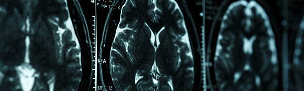
Magnetic resonance spectroscopic imaging (MRSI) performed at 7.0 T (7.0-T MRSI) could visualize pathologic findings of multiple sclerosis (MS) which were not detectable using T1- or T2-weighted magnetic resonance imaging (MRI), according to study findings published in Radiology.
Focal white matter lesions are the most detectable feature for patients with MS, however, these lesions fail to fully explain the topographical origin and severity of many clinical symptoms among patients with MS. Recent advances in 7.0-T MRSI technology have the potential to allow for more comprehensive visualization of clinical disease features.
Patients (n=65) with MS and healthy control individuals (n=20) were recruited at the Medical University of Vienna in Austria and Jessenius Faculty of Medicine in Martin in Slovakia between 2016 and 2017. Patients underwent three-dimensional T1-weighted MRI, three-dimensional T2-weighted MRI using fluid-attenuated inversion recovery, and free-induction decay MRSI at 7.0 T. Using the Expanded Disability Status Scale (EDSS), patients were stratified into low (EDSS 0-1; n=26), intermediate (EDSS 1.5-3; n=27), and advanced (EDSS ³3.5; n=12) disability groups.
The MS cohort and individuals in the control cohort were comprised 34 and 11 women and 31 and 9 men and were aged mean 34±9 and 32±7 years, respectively. Among the patient group, 61 had relapsing-remitting MS and 4 secondary progressive MS.
Normal-appearing white matter (NAWM) in the centrum semiovale was the most different between control individuals (mean, myo-inositol to N-acetylaspartate [mI/NAA] ratio, 0.56; myo-inositol to total creatine [mI/tCr] ratio, 0.78) and the patients with EDSS 0-1 (mean, mI/NAA, 0.64; P =.03 mI/tCr, 0.86; P =.02), EDSS 1.5-3 (mean, mI/NAA, 0.76; P <.001 mI/tCr, 0.95; P <.001), and ³3.5 (mean, mI/NAA, 0.75; P <.001 mI/tCr, 0.94; P=.001)
Frontal cortical gray matter (CGM) was observed to be less affected than NAWM overall, however, still differed between control individuals in patients in the cingulate cortex.
Neurometabolite alterations were also more pronounced in white matter lesions. The greatest difference was observed for mI/NAA among patients with EDSS 0-1, in which they had higher ratios for lesions (mean, 0.99) than NAWM (mean, 0.63; P <.001). A similar pattern was observed for mI/tCr among the same group (mean, 1.07 vs 0.83; P=.002), respectively.
Greater mI/NAA ratio was correlated with EDSS score in the centrum semiovale (r, 0.47; P <.001), frontal CGM (r, 0.37; P=.004), parietal NAWM (r, 0.43; P=.002), and frontal NAWM (r, 0.34; P=.01). Also, higher mI/tCr was associated with a higher EDDS score in the NAWM of centrum semiovale (r, 0.35; P=.007). The ratio between N-acetylaspartate and total creatine was negatively correlated with EDSS score in the frontal CGM (r, -0.28; P=.04) and cingulate cortex (r, -0.32; P=.02), and also the ratio between total choline to total creatine in white matter lesions (r, -0.38; P <.001).
This study was limited by the area assessed by the 7.0-T MRSI which was a single two-dimensional section measuring 8 mm.
“MR spectroscopic imaging at 7.0 T allowed in vivo visualization of multiple sclerosis pathologic findings not visible at T1- or T2-weighted MRI,” the researchers concluded.
Disclosure: Multiple authors declared affiliations with industry. Please refer to the original article for a full list of disclosures.
Reference
Heckova E, Dal-Bianco A, Stasser B, et al. Extensive Brain Pathologic Alterations Detected with 7.0-T MR Spectroscopic Imaging Associated with Disability in Multiple Sclerosis. Radiology. Published online January 4, 2022. doi:10.1148/radiol.210614
Written by: Jessica Nye, PhD